Original Article

고페리틴혈증 동반 소아다기관염증증후군의 특징

임정우1https://orcid.org/0000-0002-0227-3809, 김범준1https://orcid.org/0000-0001-5033-4568, 이수영1,2,*https://orcid.org/0000-0002-5354-3135, 정대철1,2https://orcid.org/0000-0003-0934-817X
Jung Woo Rhim1https://orcid.org/0000-0002-0227-3809, Beom Joon Kim1https://orcid.org/0000-0001-5033-4568, Soo-Young Lee1,2,*https://orcid.org/0000-0002-5354-3135, Dae Chul Jeong1,2https://orcid.org/0000-0003-0934-817X
Author Information & Copyright
1가톨릭대학교 의과대학 소아과학교실
2가톨릭대학교 의과대학 백신바이오연구소
1Department of Pediatrics, College of Medicine, The Catholic University of Korea, Seoul, Korea
2The Vaccine Bio Research Institute, College of Medicine, The Catholic University of Korea, Seoul, Korea
*Corresponding author: Soo-Young Lee, Department of Pediatrics, Bucheon St. Mary’s Hospital, The Catholic University of Korea, 327 Sosa-ro, Wonmi-gu, Bucheon, Korea, Tel: +82-32-340-2080, Fax: +82-32-340-2314, E-mail: sylee@catholic.ac.kr

ⓒ Copyright 2023 Korean Society of Kawasaki Disease. This is an Open-Access article distributed under the terms of the Creative Commons Attribution Non-Commercial License (http://creativecommons.org/licenses/by-nc/4.0/) which permits unrestricted non-commercial use, distribution, and reproduction in any medium, provided the original work is properly cited.

Received: Oct 01, 2023; Revised: Nov 17, 2023; Accepted: Dec 01, 2023

Published Online: Dec 31, 2023

Abstract

Background: Multisystem inflammatory syndrome in children (MIS-C) is a serious complication of COVID-19. Ferritin levels are used to assess severity and to predict the clinical course of various systemic inflammatory diseases that can cause cytokine storm. In this study, we investigated the clinical manifestations of patients with MIS-C and compared the characteristics of MIS-C with hyperferritinemia and MIS-C without hyperferritinemia. Methods: Medical records of 22 patients hospitalized with MIS-C from January 2020 to December 2022 were retrospectively reviewed. The characteristics of the MIS-C with hyperferritinemia group (n = 11) and the MIS-C without hyperferritinemia group (n = 11) were compared. Results: All 22 patients with MIS-C had hyperinflammation, multi-organ dysfunction, and laboratory evidence of SARS-CoV-2 infection. Compared to the MIS-C without hyperferritinemia group, the MIS-C with hyperferritinemia group had more severe systemic inflammation (e.g., thrombocytopenia, hypoalbuminemia, and two or more immunomodulators) and organ dysfunction (e.g., shock, number of involved organs, and intensive care unit admission). There were no significant differences in demographics and clinical findings between the two groups. Conclusion: We found that hyperferritinemia is an important feature of severe MIS-C in terms of systemic inflammation and organ dysfunction. Subsequent studies are needed to evaluate the clinical significance of hyperferritinemia in various Kawasaki disease (KD)-like hyperinflammatory diseases, including MIS-C and severe KD.

Keywords: Kawasaki Disease; Multisystem Inflammatory Syndrome in Children; Ferritin

서론

소아다기관염증후군(multisystem inflammatory syndrome in children, MIS-C)은 코로나바이러스감염증-19(coronavirus disease 2019, COVID-19)의 드물지만 심각한 합병증이다[1]. MIS-C의 특징은 신종 코로나바이러스(severe acute respiratory syndrome coronavirus 2, SARS-CoV-2) 감염에 의해 촉발된 전신염증과 장기부전이며, 이 세가지 요소를 증명하여 MIS-C를 진단한다(Table 1). 많은 MIS-C 환자들이 가와사키병(Kawasaki disease, KD) 진단기준에 포함되는 ‘KD-유사증상’(KD-like features)을 보여 임상적 관심을 받았다[1,2]. 실제로 MIS-C와 KD는 임상 및 검사실 소견에서 많은 유사점을 공유한다[3,4]. 하지만 인구통계학적으로 KD와는 다르게, MIS-C는 상대적으로 흑인과 히스패닉 인종에 흔하고 아시아 인종에는 드물다[5]. 국내에는 MIS-C 발생 자체가 적어서인지, MIS-C에 관한 대부분의 연구는 복수의 환자를 대상으로 하는 임상연구보다는 종설이나 증례보고 형태이다.

Table 1. CSTE/CDC definition of confirmed MIS-C case [4,13]
A patient aged < 21 years with fever (≥ 38.0°C) and clinical severity requring hospitalization
1. Systemic inflammation: CRP ≥ 3.0 mg/dL
2. Organ involvement ≥ 2/5 categories
 i) Cardiac: EF < 55%, CAAs, or Tn elevation
 ii) Mucocutaneous: skin rash, conjunctivitis, oropharyngeal inflammation, or extremity changes
 iii) Shock: clinician documentation in medical records
 iv) Gastrointestinal: abdominal pain, vomiting, or diarrhea
 v) Hematologic: platelet count < 150,000 /μL or ALC < 1,000 /μL
3. Laboratory evidence of SARS-CoV-2 infection detection of SARS-CoV-2 RNA (i.e., PCR) or SARS-CoV-2-specific antigen or antibodies up to 60 days before or during hospitalization

CSTE/CDC: The Council of State and Territorial Epidemiologists/Centers for Disease Control and Disease Prevention; MIS-C: multisystem inflammatory syndrome in children; CRP: C-reactive protein; EF: ejection fraction; CAAs: coronary artery abnormalities; Tn: troponin; ALC: absolute lymphocyte count; SARS-CoV-2: severe acute respiratory syndrome coronavirus 2.

Download Excel Table

MIS-C 환자의 조직손상과 장기부전은 선천면역(innate immunity)에 의해 매개된다고 알려져 있다[3,6]. 조절에서 벗어난 면역반응은 대식세포활성증후군(macrophage activation syndrome, MAS)의 치명적인 염증반응과 유사한 사이토카인 폭풍(cytokine storm)을 일으킨다[7,8]. 고페리틴혈증(hyperferritinemia)은 사이토카인 폭풍과 같은 심한 염증반응이 발생할 때 동반되는 검사실 소견이다[9]. 페리틴(ferritin) 수치는 염증의 중증도에 비례하여 증가하는데, 이러한 특성은 감염, 류마티스 및 혈액종양 등의 여러 종류 질환의 진단과 질병 활성도 평가에 적용되고 있다[6,10]. ‘중증(severe) COVID-19’ 성인 환자 대상의 연구에서, 생존(survivor) 환자군에 비해 비생존(non-survivor) 환자군에서 페리틴 수치의 급격한 증가가 확인되었다[11]. 이는 MIS-C 환자에서도 페리틴 검사가 단순히 전신염증을 입증하는 것뿐만 아니라, 임상적으로 좀더 다양한 측면에서 활용될 수 있음을 의미한다.

본 연구에서는 국내 MIS-C 환자의 임상양상을 조사하고, 고페리틴혈증 동반 MIS-C 환자와 고페리틴혈증이 없는 MIS-C 환자의 특징을 비교하여, MIS-C 환자에서 페리틴 검사의 의의를 확인하려고 한다.

대상과 방법

1. 대상

2020년 1월부터 2022년 12월까지(COVID-19 팬데믹 3년) 가톨릭의대 4개 병원(서울성모병원, 은평성모병원, 부천성모병원, 대전성모병원) 소아청소년과에 MIS-C로 입원한 환자의 의무기록을 후향적으로 조사한 연구[12]이다. MIS-C 진단코드(U10.9)의 환자 중 2023년의 새로운 사례정의(Table 1)를 만족하면서 입원기간 동안 MIS-C에 대한 검사(페리틴 포함)를 시행받고 치료를 위해 정맥주사 면역글로불린(intravenous immunoglobulin, IVIG) 혹은 정맥주사 스테로이드(intravenous methylprednisolone, IVMP) 등, 한가지 이상의 면역조절제를 투여받은 환자를 대상으로 선정하였다. MIS-C 사례정의를 만족하지 못하거나, MIS-C에 대한 검사와 치료 미시행 혹은 이에 대한 기록 누락 환자, MIS-C로 2회 이상 입원한 환자 그리고 치료 도중에 다른 병원으로 전원된 환자는 제외시켰다. 대상환자는 페리틴 수치를 토대로, ‘고페리틴혈증(페리틴 > 500 ng/mL) 동반 MIS-C군’과 ‘고페리틴혈증 없는 MIS-C군’으로 양분하였다.

2. 정의

MIS-C 진단에는 미국의 국가 및 영토 역학위원회 및 질병통제예방센터(Council of State and Territorial Epidemiologists/Centers for Disease Control and Disease Prevention, CSTE/CDC)에서 새롭게 제정한 2023년 MIS-C 사례정의[13]를 이용했다. KD는 미국심장학회(American Heart Association, AHA) 기준[14]에 따라 완전형(complete)과 불완전형(incomplete)으로 진단하였다. 일차치료의 실패(즉, IVIG 저항성)는 첫번째 IVIG ± IVMP 치료 종결 36시간 후에 발열이 지속되어 두번째 면역조절제가 투여된 경우로 정의하였다[15]. 관상동맥 이상(coronary artery abnormalities, CAAs)은 AHA의 Z score 기준에 따라, Z score 2.0 이상은 관상동맥 확장으로, Z score 2.5 이상은 관상동맥류로 정의하였다[14].

3. 관찰항목

임상적 항목으로 연령, 성별, 입원기간, 입원시 활력증후, 발열 기간, KD-유사증상(발진, 결막염, 입술과 입 안의 염증, 손발의 변화, 경부림프절 비대) 및 장기부전의 양상(심장, 점막피부, 쇼크, 소화기, 혈액학적, 그 밖의 주요 장기)이 포함되었다. 검사실 항목으로 혈색소, 백혈구, 절대 림프구(absolute lymphocyte count, ALC), 혈소판, 적혈구 침강속도(erythrocyte sedimentation rate, ESR), C-반응단백(C-reactive protein, CRP), aspartate aminotransferase (AST), alanine aminotransferase(ALT), 알부민, N-terminal pro-brain natriuretic peptide(NT-proBNP), 트로포닌(troponin), 페리틴, 중성지방, 섬유소원(fibrinogen), 응고이상(international normalized ratio, INR) 및 농뇨(pyuria)를 조사하였고, 별도로 2016년 MAS 진단기준[16]의 충족 여부를 평가하였다. 미생물학적 검사(일반 바이러스와 세균 배양검사, polymerase chain reaction (PCR) 및 혈청학)와 심장 초음파 검사 결과도 확인하였다.

SARS-CoV-2 감염 증명을 위해, 과거 COVID-19 이환력과 당시 시행한 검사 결과 그리고 입원시 SARS-CoV-2에 대한 PCR, 항원 및 항체 검사 결과를 확인하였다. 치료와 임상경과의 평가를 위해, 일차 치료약제, 치료 실패에 대한 추가적 치료약제, 경험적 항생제, 혈압상승제, 중환자실 입실 및 사망 여부를 조사하였다. 또한 KD 환자와 비교한 MIS-C 환자 발생현황을 파악하기 위해, 연구기간 3년 동안 KD로 입원한 환자 수도 조사하였다.

4. 통계분석

범주형 자료는 빈도와 퍼센트로, 연속형 자료는 중앙값과 범위로 나타냈다. 범주형 자료의 비교는 Fisher 정확 검정을, 연속형 자료의 비교는 Mann-Whitney U 검정을 이용했다. 모든 통계분석은 양측(two-tailed) 검정으로, P < 0.05를 통계적으로 유의한 것으로 간주하였다. 통계 프로그램은 IBM SPSS Statistics software Version 24.0(IBM, Armonk, NY, USA)을 이용했다.

결과

1. 전체 MIS-C 환자 수

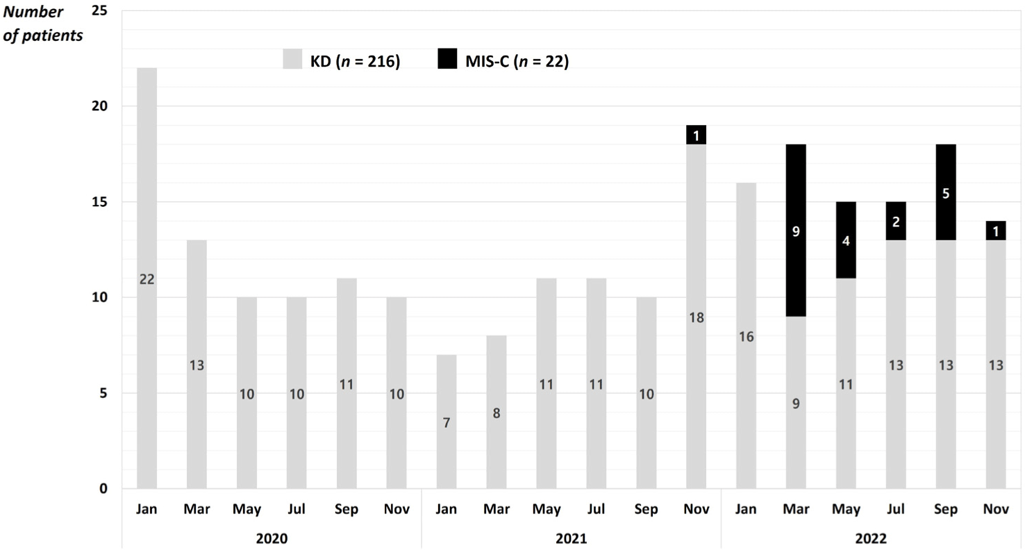
사례정의를 만족하지 못한 환자 4명과 전원된 환자 1명을 제외하고, 총 22명의 MIS-C 환자가 연구대상으로 선정되었다. 이중 11명(50.0%)은 고페리틴혈증 동반 MIS-C군으로, 나머지 11명(50.0%)은 고페리틴혈증 없는 MIS-C군으로 할당되었다. 모든 환자들(100.0%)은 (1) 전신염증(CRP ≥ 3.0 mg/dL)과 (2) 두 가지 이상 장기의 기능부전을 나타냈고, (3) 미생물학적으로 SARS-CoV-2 감염이 증명되었다. 연도별 MIS-C 입원 환자수는 2020년 0명(0.0%), 2021년 1명(4.6%), 2022년 21명(95.4%)이며, Fig. 1에 KD 환자와 비교한 MIS-C 환자 발생현황을 나타냈다. MIS-C/KD 비율은 0.10(22/216)으로, 미국 MIS-C/KD 비율 1.66(31.6/19.0)의 1/16 수준이다[17].

kd-1-2-16-g1
Fig. 1. The incidence of MIS-C and KD from January 2020 to December 2022 at four university hospitals in Korea. The incidence of MIS-C compared to KD was 0.10 (22/216), which was less than 1/16 of that in the United States (1.66; 31.6/19.0) [17]. MIS-C: multisystem inflammatory syndrome in children; KD: Kawasaki disease.
Download Original Figure
2. 임상적 소견과 장기부전

전체 환자 연령의 중앙값은 9.3세(0.9–16.0)였고, 남녀 성비는 1:1이었다. 입원기간의 중앙값은 10.0일(4.0–18.0), 입원 전 발열기간의 중앙값은 5.0일(3.0–15.0)이었다. KD-유사증상 중에는 발진(77.3%)과 결막염(81.8%)이 흔하였고, 입술과 입 안의 염증, 손발의 변화, 경부림프절 비대는 각각 45.5%, 59.1%, 68.2%의 환자에서 관찰되었다. KD 진단기준을 만족한 환자는 19명(86.4%)이며, 이중 11명(50.0%)은 완전형 KD였다. 장기부전 측면에서, 심장 증상 12명(54.5%), 점막피부 증상 21명(95.5%), 쇼크 증상 6명(27.3%), 소화기 증상 20명(90.9%), 혈액학적 증상 17명(77.3%), 그 외 호흡기, 신경 혹은 신장 증상은 14명(63.6%)에서 관찰되었다.

Table 2에는 고페리틴혈증 동반 MIS-C군과 고페리틴혈증이 없는 MIS-C군의 임상적 특징과 장기침범 양상을 비교하였다. 연령, 성비, KD-유사증상을 포함한 임상적 특징에는 두 군간 유의한 차이가 없었고, 장기침범 양상에는 쇼크 증상의 빈도(54.5%)와 침범된 장기의 중앙값(4.0)이 고페리틴혈증 없는 MIS-C군보다 고페리틴혈증 동반 MIS-C군에서 높았다(P < 0.05).

Table 2. Clinical characteristics and organ involvement of MIS-C with or without hyperferritinemia
MIS-C with hyperferritinemia (n = 11) MIS-C without hyperferritinemia (n = 11) P value
Clinical
 Age, year 9.1 (4.1–16.0) 9.5 (0.9–12.0) 0.669
 Male sex 5 (45.5) 6 (54.5) 0.670
 Length of hospital stays, day 10.0 (5.0–18.0) 6.0 (4.0–15.0) 0.072
 Fever duration, day 5.0 (3.0–15.0) 5.0 (3.0–10.0) 0.917
 Skin rash 7 (63.6) 10 (90.9) 0.311
 Met KD criteria 10 (90.9) 9 (81.8) 1.000
 Complete KD 6 (54.5) 5 (45.5) 1.000
Organ involvement (see Table 1)
 Cardiac involvement 8 (72.7) 4 (36.4) 0.198
 Mucocutaneous involvement 11 (100.0) 10 (90.9) 1.000
 Shock 6 (54.5) 0 (0.0) 0.012
 Gastrointestinal involvement 11 (100.0) 9 (81.8) 0.476
 Hematologic involvement 10 (90.9) 7 (63.6) 0.311
 Others: respiratory, neurologic, or renal1) 8 (72.7) 6 (54.5) 0.659
 Number of organs involved 4.0 (2.0–5.0) 3.0 (2.0–4.0) 0.002

Data are presented as frequency (%) or median (range). MIS-C: multisystem inflammatory syndrome in children; KD: Kawasaki disease.

1) The Council of State and Territorial Epidemiologists/Centers for Disease Control and Disease Prevention (CSTE/CDC) case definition includes five types of organ involvement (i.e., cardiac, mucocutaneous, shock, gastrointestinal, and hematologic). Other organ dysfunction (e.g., respiratory, neurologic, renal, or musculoskeletal) is not included in the CSTE/CSC case definition.

Download Excel Table
3. 검사실 소견

전체 환자의 검사실 소견은 다음과 같다: 혈색소 12.1 g/dL (9.6–14.9), 백혈구 7,200 /μL (2,350–20,390), ALC 937 /μL (198–6,890), 혈소판 130 × 103 /μL (62–431), ESR 26 mm/hr (2–120), CRP 10.1 mg/dL (3.2–25.5; 정상 < 5.0 mg/L), AST 55 U/L (33–579), ALT 54 U/L (13–232), 알부민 2.9 g/dL (2.4–4.1), NT-proBNP 1,464 pg/dL (299–10,364), 페리틴 566 ng/mL (158–10,364), 트로포닌 상승 8명(36.4%), 중성지방 혹은 섬유소원 이상 17명(77.3%), 응고이상 15명(68.2%) 및 2016년 MAS 진단기준 만족 11명(50%)이었다.

Table 3에는 고페리틴혈증 동반 MIS-C군과 고페리틴혈증 없는 MIS-C군의 검사실 특징을 비교하였다. 고페리틴혈증 없는 MIS-C군과 비교하여, 고페리틴혈증 동반 MIS-C군에서 혈소판 수치와 알부민 수치는 낮았고 페리틴 수치는 높았으며, 트로포닌 상승 환자의 비율과 2016년 MAS 진단기준 만족 환자의 비율이 높았다(P < 0.05).

Table 3. Laboratory characteristics of MIS-C with or without hyperferritinemia
MIS-C with hyperferritinemia (n = 11) MIS-C without hyperferritinemia (n = 11) P value
Hemoglobin, g/dL 12.1 (9.6–14.9) 12.1 (10.5–13.2) 0.693
Total WBC count, μL 7,110 (2,350–20,390) 7,290 (2,940–14,660) 0.718
ALC, μL 854 (198–2,260) 1,020 (414–6,890) 0.450
Platelet count, 103 /μL 103 (62–431) 181 (103–389) 0.010
ESR, mm/h 22 (2–120) 36 (11–105) 0.211
CRP, mg/dL (normal < 0.5) 13.8 (3.2–25.5) 5.1 (3.3–9.4) 0.130
AST, U/L 52 (40–579) 57 (33-94) 0.843
ALT, U/L 54 (34–232) 61 (13–120) 0.412
Albumin, g/dL 2.8 (2.5–3.2) 3.4 (2.3–4.1) 0.048
NT-proBNP, pg/dL 1,044 (421–23,858) 1,676 (299–3,662) 0.577
Ferritin, ng/mL 1,145 (690–10,364) 286 (148–443) < 0.001
Tn elevation 7 (63.6) 1 (11.1) 0.028
Abnormal TG or fibrinogen1) 8 (72.7) 9 (81.8) 1.000
Coagulopathy: INR > 1.1 7 (63.6) 8 (72.7) 1.000
Pyuria: ≥ 10 WBC/hpf 5 (45.4) 2 (18.2) 0.361
Met 2016 MAS criteria2) 11 (100.0) 0.0 (0.0) < 0.001

Data are presented as frequency (%) or median (range). MIS-C: multisystem inflammatory syndrome in children; WBC: white blood cell; ALC: absolute lymphocyte count; ESR: erythrocyte sedimentation rate; CRP: C-reactive protein; AST: aspartate transaminase; ALT: alanine transaminase; NT-proBNP: N-terminal pro-brain natriuretic peptide; Tn: troponin; TG: triglyceride; INR: international normalized ratio; hpf: high-power field; MAS: macrophage activation syndrome.

1) TG >156 mg/dL or fibrinogen ≤360 mg/dL.

2) MAS is diagnosed when ferritin > 684 ng/mL and 2 of 4 criteria (platelets ≤ 181,000/μL, AST > 48 U/L, TG > 156 mg/dL, or fibrinogen ≤ 360 mg/dL) [16].

Download Excel Table
4. 치료와 임상경과

MIS-C에 대한 초기 치료에 IVIG 투여 받은 환자는 19명(86.4%)이었다. 일차치료 실패(IVIG 저항성) 환자는 7명(31.8%)이었고, 추가적 IVIG는 4명(18.2%), 추가적 IVMP는 7명(31.8%), 생물학적 제제(anakinra)는 1명(4.5%)에서 시행되었다. 두 가지 이상의 면역조절제 치료받은 환자는 17명(77.3%)이었다. 경험적 항생제는 19명(86.4%), 혈압상승제는 5명(22.7%) 및 중환자실 입실은 5명(22.7%)에서 시행되었다. CAAs은 5명(22.7%)에서 확인되었으며, 사망한 환자(0.0%)는 없었다.

Table 4에는 고페리틴혈증 동반 MIS-C군과 고페리틴혈증 없는 MIS-C군의 치료와 임상경과를 비교하였다. 고페리틴혈증이 없는 MIS-C군과 비교하여, 고페리틴혈증 동반 MIS-C군에서는 두 가지 이상의 면역조절제 치료받은 환자의 비율, 혈압상승제 투여 환자의 비율 및 중환자실 입실 환자의 비율이 높았다(P < 0.05).

Table 4. Treatment and outcomes of MIS-C with or without hyperferritinemia
MIS-C with hyperferritinemia (n = 11) MIS-C without hyperferritinemia (n = 11) P value
Initial IVIG 11 (100.0) 8 (72.7) 0.214
Initial treatment failure (IVIG resistance) 5 (45.5) 2 (18.2) 0.361
Second or third IVIG 3 (27.3) 1 (9.1) 0.586
Second or third IVMP 5 (45.5) 2 (18.2) 0.361
Biologics (anakinra) 1 (9.1) 0 (0.0) 1.000
Two or more immunomodulators1) 11 (100.0) 6 (54.5) 0.035
Empirical antibiotics 11 (100.0) 8 (72.7) 0.214
Vasoactive drugs 5 (45.5) 0 (0.0) 0.035
ICU admission 5 (45.5) 0 (0.0) 0.035
Coronary complications 2 (18.2) 3 (27.3) 1.000
Mortality 0 (0.0) (0.0) 1.000

Data are presented as frequency (%). MIS-C: multisystem inflammatory syndrome in children; IVIG: intravenous immunoglobulin; IVMP: intravenous methylprednisolone; ICU: intensive care unit.

1) Immunomodulators include IVIG, IVMP, and anakinra.

Download Excel Table

고찰

본 연구에서 COVID-19 팬데믹 기간 동안 국내 병원에 입원한 MIS-C 환자 22명의 특징을 조사하였다. 전체 MIS-C 환자 중 11명(50%)에서 고페리틴혈증이 관찰되었다. 고페리틴혈증 동반 MIS-C군(n = 11)과 고페리틴혈증 없는 MIS-C군(n = 11)의 비교할 때, 인구학적 특징과 임상적 소견에는 유의한 차이가 없었고, 장기침범 양상과 검사실 소견에는 고페리틴혈증 동반 MIS-C군에서 좀더 심한 양상을 보였다. 치료적 측면에서도 고페리틴혈증 동반 MIS-C군에서 두 가지 이상의 면역조절제와 혈압상승제 치료 그리고 중환자실 입실이 필요했던 환자의 비율이 높았다.

소아 COVID-19는 성인에 비해 임상경과가 심하지 않다고 알려져 있다[1,2]. 하지만, 2020년 4월 이탈리아에서 Verdoni et al.[3]은 SARS-CoV-2 감염 후 심한 전신염증, 쇼크 증상을 포함한 장기침범 그리고 KD 유사증상을 보이는 소아 및 청소년을 보고하였다. 이 심각한 COVID-19 합병증은 영국, 스페인, 프랑스, 미국, 인도, 브라질 등에서도 보고되었고 ‘COVID-19 연관 소아다기관염증증후군(MIS-C)’이라 명명되었다[4]. Verdoni et al.[3]의 MIS-C 환자(n = 10)는 모두 KD 진단기준을 만족하였고 50%는 고페리틴혈증(> 500 ng/mL)을 보였으며 80%는 두 가지 이상의 면역조절제, 20%는 혈압상승제 치료를 받았고 다행히 모두 회복되었다. 이는 전반적으로 본 연구와 유사한 결과이다. Feldstein et al.[18]의 연구(n = 186)에서 KD 진단기준 만족 비율 40%, 고페리틴혈증 61%, 추가적 면역조절제 69%, 혈압상승제 48% 및 사망률 2%(4/186)를 보고하였는데, 이는 본 연구에 비해 KD 진단기준 만족 비율은 낮고 검사실 소견, 치료양식 및 임상경과는 좀 더 심한 양상이다.

MIS-C는 임상적 특징과 중증도에 따라 분류할 수 있다. Table 5에는 MIS-C 사례 보고가 급증했던 2020년 8월 미국 질병통제예방센터(CDC)가 제안한 분류법(class 1, 2 및 3)을 요약하였다[19,20]. Class 1은 급성 COVID-19의 호흡기 증상과 KD-유사증상의 빈도는 낮고 심한 검사실 소견과 더 많은 장기의 침범을 특징으로 하는 ‘전형적(classic) MIS-C’로 전체 환자의 약 35%(n = 203)를 차지한다. Class 2는 성인의 중증 COVID-19와 유사한 양상으로, 대부분 호흡기 증상을 보이며 SARS-CoV-2 PCR에 대한 높은 양성률을 보이는 ‘급성(acute) COVID-19 MIS-C’로 전체 환자의 약 30%(n = 169)를 차지한다. Class 3는 class 1과 2에 비해 KD-유사증상은 흔하지만 전신염증과 장기침범의 정도는 심하지 않은 ‘KD-유사(KD-like) MIS-C’로, 전체 환자의 약 35%(n = 198)를 차지한다. 본 연구의 MIS-C 환자들은 연령 중앙값, 장기침범의 정도, 치료 및 경과는 class 1와 유사하였고 KD-유사증상의 빈도는 class 3과 유사하였다(Table 5). 국외 연구[18-20]와 가장 큰 차이점은 고페리틴혈증이 동반된 심한 MIS-C 환자에서 KD-유사증상의 빈도가 높았다는 것이다. 이러한 차이는 소규모 연구라서 나타난 현상일 수 있겠으나, KD의 발생률은 높고 MIS-C의 발생률은 낮은 아시아 인종의 특징일 수도 있다[21].

Table 5. MIS-C groups (Class 1, 2, and 3) reported to the CDC [19] and their comparison with this study
Class 1, n = 203 (classic MIS-C) Class 2, n = 169 (acute COVID-19) Class 3, n = 198 (KD-like MIS-C) This study, n = 22
Clinical
 Age, years 9 10 6 9
 Male sex, % 57.1 52.1 56.6 50.0
 Asian race, % 0.6 2.3 5.6 100.0
 Complete KD, % 4.9 3.0 6.6 50.0
 PCR (–) & Ab (+)1), % 68.0 0.0 63.1 40.9
Organ involvement, %
 Cardiac involvement 55.3 46.0 21.0 54.5
 Mucocutaneous involvement 76.8 51.5 81.3 95.5
 Shock 75.9 28.4 0.0 27.3
 Gastrointestinal involvement 97.5 86.4 87.9 90.9
 Thrombocytopenia 41.4 26.6 23.7 59.1
Treatment and outcomes, %
 IVIG 87.9 62.7 87.5 86.4
 ICU admission 84.2 62.1 44.4 22.7
 Ventilator care (ARDS) 6.9 10.1 1.5 4.5
 Coronary complications 21.1 15.8 18.2 22.7
 Mortality 0.5 5.3 0.0 0.0

1) PCR (–) & Ab (+) indicate that there is no more virus but a serious post-infectious inflammatory response, which is the key pathogenesis of MIS-C.

MIS-C: multisystem inflammatory syndrome in children; CDC: Centers for Disease Control and Disease Prevention; COVID-19: coronavirus disease 2019; KD: Kawasaki disease; PCR: polymerase chain reaction; Ab: antibodies; IVIG: intravenous immunoglobulin; ICU: intensive care unit; ARDS: acute respiratory distress syndrome.

Download Excel Table

KD-유사증상은 장기침범의 한 형태로 MIS-C 사례 정의에 포함되는 MIS-C의 흥미로운 특징이다[13]. 하지만 KD-유사증상은 질병 초기에 나타나지 않기도 하고 일부(~25%) 환자에서는 질병의 전 과정 동안 전혀 나타내지 않을 수 있어, MIS-C 진단 지연의 원인이 되기도 한다[18,22]. 국내 첫 MIS-C 소아 환자[23]의 경우, 초기에 KD-유사증상 없이 발열과 복통, 쇼크 증상을 보여 위장관계 감염에 의한 패혈증 쇼크로 오인되기도 하였다. 발진, 결막염 및 입술과 입 안의 염증은 입원 6일째 나타났고 혈청학에서 SARS-CoV-2 감염이 입증되어 최종적으로 MIS-C로 진단되었다. 따라서 KD-유사증상 유무와 무관하게, 원인이 설명되지 않는 심부전, 복통 및 쇼크 증상을 보이는 환자에서 MIS-C의 동반 가능성을 고려해야 하는 것이 중요하다[5]. 페리틴 수치는 모호한 의학적 상황에서 MIS-C 감별진단을 위해 필수적으로 확인해야 하는 검사항목이다[4].

페리틴은 철(iron) 저장 단백질로 우리 몸에서 철분 대사작용뿐만 아니라 중요한 면역학적 기능을 수행한다. 고페리틴혈증에 관한 Senjo et al.[9]의 연구(n = 1,394)에서, 페리틴 수치 500 ng/mL 이상인 질환은 감염 질환(44.8%), 고형장기 종양(26.3%), 간기능 이상(20.3%), 신장 부전(20.2%), 혈액 악성종양(12.0%), 철분대사 이상(6.9%), 류마티스/염증 질환(6.3%), MAS (1.4%) 등의 순으로 보고했다. 페리틴 중앙값이 높은 질환은 MAS (44,901 ng/mL), 혈액 및 고형장기 종양(1,165–1,330 ng/mL) 그리고 류마티스/염증 질환(1,142 ng/mL) 등의 순이었다. 페리틴 자체는 비특이적 급성기 반응 물질(acute phase reactant)이지만, 숙주 염증의 중증도에 따라 구별되는 특성을 보이기에, 류마티스 질환이나 혈액종양 질환뿐만 아니라 심한 전신염증을 유발하는 다양한 임상적 상황에서 페리틴을 활용할 수 있다[6,8].

최근 KD 연구에서 페리틴 평가의 유용성이 제시되었다[2428]. KD 환자에서 페리틴 검사를 통해, KD와 일반 열성질환(nonspecific febrile illnesses)을 구분하고[24] KD 환자의 IVIG 저항성과 CAAs 발생 가능성을 예측하며[25], 드물지만(1%–2%) 심각한 경과의 KD 합병 MAS (KD complicated with MAS)을 조기에 진단할 수 있다[26,28]. 이러한 임상적 유용성을 토대로, 페리틴 검사가 치료불응 KD와 같은 심한 형태의 KD 환자에서는 필수 검사항목에 포함되어야 한다고 제안되었다[10,15]. 유사한 이유에서, 페리틴 검사는 일반 COVID-19 소아 환자에서 필수가 아니겠지만 MIS-C 환자와 중증 COVID-19 환자에서는 필수적으로 시행되어야 한다고 여겨진다. 이미 많은 의료기관에서 MIS-C 환자에서 페리틴 검사를 시행하지만 진단시 심한 전신염증 확인을 위한 검사뿐만 아니라, 연속적으로 페리틴 수치를 확인하여 치료반응을 평가하고 예후를 예측할 수 있다. 다양한 소아 질환에서 시행된 페리틴에 대한 연구를 종합하여 각 질환들을 페리틴의 평균값 혹은 중앙값 순서로 나열하면, 일반 열성질환(예, 아데노바이러스 감염; 107 ng/mL 미만)[24] < 전형적 KD (121 ng/mL 이상)[24] < 치료불응 KD (165 ng/mL 이상)[25] < MIS-C (566 ng/mL)[12] < KD 쇼크증후군 (756 ng/mL)[12] < KD 합병 MAS (3,490 ng/mL)[27] 순이다. 즉, KD-유사증상(발진, 결막염, 입술과 입 안의 염증 등)을 나타낼 수 있는 다양한 과염증 질환들(KD-like hyperinflammatory diseases)을 MIS-C와 구분할 때 페리틴 검사가 활용 가능하다[5].

본 연구는 대상환자가 소규모이며 제한된 지역 병원에 입원한 MIS-C 환자의 의무기록을 후향적으로 조사하였기에 그 결과를 특정 지역이나 국가 전체의 자료로 대체하기에는 한계가 있다. 또한, 진단코드를 이용하여 대상환자를 선별하였기에 실제 환자가 누락되었을 수 있고 그에 따라 MIS-C 발생빈도와 임상양상이 과소 평가되었을 수 있다. 하지만 본 연구에서 아직 많은 자료가 필요한 국내 MIS-C의 대략적 발생현황과 임상양상을 조사하였고 고페리틴혈증이 전신염증과 장기침범 측면에서 심한 MIS-C의 특징이라는 것을 확인하였다. COVID-19 팬데믹 종결이 선언된 시점에서 MIS-C의 역학적, 임상적 변화를 확인하기 위한 추가적 연구가 요구된다.

Conflict of Interest

No potential conflict of interest relevant to this article was reported.

Funding

No funding source relevant to this article was reported.

Acknowledgements

Not applicable.

Authors’ Contributions

Conceptualization: Rhim JW.

Data curation: Rhim JW, Kim BJ.

Formal analysis: Lee SY.

Methodology: Kim BJ, Jeong DC.

Software: Rhim JW.

Validation: Lee SY.

Investigation: Jeong DC.

Writing - original draft: Rhim JW.

Writing - review & editing: Rhim JW, Kim BJ, Lee SY, Jeong DC.

Ethics Approval

This study was approved by the Institutional Review Board of Bucheon St. Mary’s Hospital, The Catholic University of Korea (IRB No. HC23RISI0099). The requirement of obtaining informed consent was waived by the board.

References

1.

Sharma C, Ganigara M, Galeotti C, Burns J, Berganza FM, Hayes DA, et al. Multisystem inflammatory syndrome in children and Kawasaki disease: a critical comparison. Nat Rev Rheumatol. 2021; 17:731-48

2.

Kwak JH, Lee SY, Choi JW. Clinical features, diagnosis, and outcomes of multisystem inflammatory syndrome in children associated with coronavirus disease 2019. Clin Exp Pediatr. 2021; 64:68-75

3.

Verdoni L, Mazza A, Gervasoni A, Martelli L, Ruggeri M, Ciuffreda M, et al. An outbreak of severe Kawasaki-like disease at the Italian epicentre of the SARS-CoV-2 epidemic: an observational cohort study. Lancet. 2020; 395:1771-8

4.

Shin EJ. Kawasaki disease and multisystem inflammatory syndrome in children. Kawasaki Dis. 2023; 1e4

5.

Lee S, Kim D, Kim BJ, Rhim JW, Lee SY, Jeong DC. Comparison of COVID-19-associated multisystem inflammatory syndrome in children (MIS-C) and Kawasaki disease shock syndrome: case reports and literature review. J Rheum Dis. 2023; 30:272-7

6.

Mahroum N, Alghory A, Kiyak Z, Alwani A, Seida R, Alrais M, et al. Ferritin - from iron, through inflammation and autoimmunity, to COVID-19. J Autoimmun. 2022; 126:102778

7.

Mehta P, McAuley DF, Brown M, Sanchez E, Tattersall RS, Manson JJ. COVID-19: consider cytokine storm syndromes and immunosuppression. Lancet. 2020; 395:1033-4

8.

Cheng L, Li H, Li L, Liu C, Yan S, Chen H, et al. Ferritin in the coronavirus disease 2019 (COVID-19): a systematic review and meta-analysis. J Clin Lab Anal. 2020; 34e23618

9.

Senjo H, Higuchi T, Okada S, Takahashi O. Hyperferritinemia: causes and significance in a general hospital. Hematology. 2018; 23:817-22

10.

Roh DE, Kwon JE, Choi HJ, Kim YH. Importance of serum ferritin level for early diagnosis and differentiation in patients with Kawasaki disease with macrophage activation syndrome. Children (Basel). 2021; 8:269

11.

Zhou F, Yu T, Du R, Fan G, Liu Y, Liu Z, et al. Clinical course and risk factors for mortality of adult inpatients with COVID-19 in Wuhan, China: a retrospective cohort study. Lancet. 2020; 395:1054-62

12.

Lee J, Kim BJ, Cho KS, Rhim JW, Lee SY, Jeong DC. Similarities and differences between multisystem inflammatory syndrome in children (MIS-C) and Kawasaki disease shock syndrome. Children (Basel). 2023; 10:1527

13.

Melgar M, Lee EH, Miller AD, Lim S, Brown CM, Yousaf AR, et al. Council of State and Territorial Epidemiologists/CDC surveillance case definition for multisystem inflammatory syndrome in children associated with SARS-CoV-2 infection—United States. MMWR Recomm. Rep. 2022; 71:1-14

14.

McCrindle BW, Rowley AH, Newburger JW, Burns JC, Bolger AF, Gewitz M, et al. Diagnosis, treatment, and long-term management of Kawasaki disease: a scientific statement for health professionals from the American Heart Association. Circulation. 2017; 135:e927-99

15.

Rhee S, Kim D, Cho K, Rhim JW, Lee SY, Jeong DC. Under-recognized macrophage activation syndrome in refractory Kawasaki disease: a wolf in sheep’s clothing. Children (Basel). 2022; 9:1588

16.

Ravelli A, Minoia F, Davì S, Horne AC, Bovis F, Pistorio A, et al. 2016 Classification criteria for macrophage activation syndrome complicating systemic juvenile idiopathic arthritis: a European league against rheumatism/American College of Rheumatology/Paediatric Rheumatology International Trials Organisation Collaborative Initiative. Ann Rheum Dis. 2016; 75:481-9

17.

Payne AB, Gilani Z, Godfred-Cato S, Belay ED, Feldstein LR, Patel MM, et al. Incidence of multisystem inflammatory syndrome in children among US persons infected with SARS-CoV-2. JAMA Netw Open. 2021; 4e2116420.

18.

Feldstein LR, Rose EB, Horwitz SM, Collins JP, Newhams MM, Son MBF, et al. Multisystem inflammatory syndrome in U.S. children and adolescents. N Engl J Med. 2020; 383:334-46

19.

Godfred-Cato S, Bryant B, Leung J, Oster ME, Conklin L, Abrams J, et al. COVID-19-associated multisystem inflammatory syndrome in children - United States, March–July 2020. MMWR Morb Mortal Wkly Rep. 2020; 69:1074-80

20.

Rowley AH, Shulman ST, Arditi M. Immune pathogenesis of COVID-19-related multisystem inflammatory syndrome in children. J Clin Invest. 2020; 130:5619-21

21.

You SD, Kim JH, You J. Clinical characteristics and short-term outcomes of multisystem inflammatory syndrome in a country with a high prevalence of KD. Front Pediatr. 2023; 11:1088529

22.

Ludwikowska KM, Okarska-Napierała M, Dudek N, Tracewski P, Kusa J, Piwoński KP, et al. Distinct characteristics of multisystem inflammatory syndrome in children in Poland. Sci Rep. 2021; 11:23562

23.

Kim H, Shim JY, Ko JH, Yang A, Shim JW, Kim DS, et al. Multisystem inflammatory syndrome in children related to COVID-19: the first case in Korea. J Korean Med Sci. 2020; 35:e391-8

24.

Kim SH, Song ES, Yoon S, Eom GH, Kang G, Cho YK. Serum ferritin as a diagnostic biomarker for Kawasaki disease. Ann Lab Med. 2021; 41:318-22

25.

Yamamoto N, Sato K, Hoshina T, Kojiro M, Kusuhara K. Utility of ferritin as a predictor of the patients with Kawasaki disease refractory to intravenous immunoglobulin therapy. Mod Rheumatol. 2015; 25:898-902

26.

Han SB, Lee SY. Macrophage activation syndrome in children with Kawasaki disease: diagnostic and therapeutic approaches. World J Pediatr. 2020; 16:566-74

27.

García-Pavón S, Yamazaki-Nakashimada MA, Báez M, Borjas-Aguilar KL, Murata C. Kawasaki disease complicated with macrophage activation syndrome: a systematic review. J Pediatr Hematol Oncol. 2017; 39:445-51

28.

Kim BJ, Kim D, Cho K, Rhim JW, Lee SY, Jeong DC. Kawasaki disease shock syndrome and macrophage activation syndrome: a case report. Kawasaki Dis. 2023; 1e11